Bone Problems: 4 Key Examples of Musculoskeletal Disorders

An estimated 1.71 billion people across the world live with musculoskeletal disorders. It is considered one of the biggest reasons for disability. If you are sick and tired of bone problems, you may be looking for several key examples of musculoskeletal symptoms and disorders to watch out for. 

You can find out more here. Keep reading to learn more about common symptoms of musculoskeletal disorders and conditions. 

1. Rheumatoid Arthritis 

Rheumatoid arthritis (RA) is a condition marked by chronic inflammation of the joints. It differs from arthritis as RA affects the immune system.  With RA, the body attacks its own tissues, which may cause flu-like symptoms. 

RA may damage more parts of the body aside from the joints. Using many treatments, including medication and physical therapy, is vital for managing flareups. 

2. Carpal Tunnel Syndrome 

Carpal tunnel syndrome (CTS) is a musculoskeletal disorder that happens when the nerve that runs from the palm down the forearm is put under pressure. It may appear as muscle weakness, tingling, or pain. Injury to the wrist or repetitive movements may trigger CTS. 

Improved posture, stretching, and resting or alternating between activities can help prevent this disorder. However, after developing CTS treatment, such as splinting, medication, or physical therapy may be necessary. 

3. Fibromylagia 

Fibromyalgia is a common autoimmune condition impacting around 2% of adults. It is characterized by widespread chronic pain, difficulty sleeping, excessive daytime fatigue, brain fog, depression, and anxiety. The unpredictable and disabling pain of fibromyalgia can make daily functioning difficult. 

Strengthening exercises are an important treatment for fibromyalgia. Regular physical activity can help manage pain and the other symptoms of this condition.

4. Back Pain 

“Back pain” is not a term referring to any specific musculoskeletal disorders themselves. It is one of the most common types of chronic pain experienced by adults at one or more points in their lives. Back pain can be caused by arthritis, conditions affecting the spinal disks, physical injury, structural abnormalities, genetics, poor posture, or aging. Diagnosis is not always the most essential part of relieving pain in the back.

Many types of lower back pain can be helped with movement-based therapies. Exercises that focus on strengthening and stretching the muscles in the core and legs can work to relieve and prevent pain. It is crucial to consult a doctor or physical therapist before starting a new exercise or stretching routine on your own to avoid injury or strain. 

Find Relief From Your Musculoskeletal Disorders Now

A great way to ease the pain that comes with musculoskeletal disorders like arthritis, carpal tunnel, tendonitis, or fibromyalgia is with physical therapy or therapeutic massage. We will develop a personalized treatment plan for you to help in restoring mobility and strength. As experts in our field, we strive to assist all of our patients to live healthy, pain-free lives. 

If you want to learn more about our physical therapy or therapeutic massage programs for musculoskeletal disorders and symptoms, contact us today.

If your back hurts, a diagnosis may not be the best path

Low back pain is jarringly common. About one-half of all working Americans will experience symptoms at least once every year, and roughly 31 million are affected by it at any given point in time. So if you find it appropriate to place yourself in this category, you’d have an abundance in company.

Dealing with low back pain can be troublesome and place a strain on everyday life. Typical movements like bending over to pick something off the ground or twisting your torso when looking to the side might suddenly give you pause and make you less mobile in the process. This development naturally leads to frustration and often shifts to a focus on one main question: “what’s causing this pain?”

As a result, many patients with low back pain start to place a particularly strong—and sometimes unhealthy—emphasis on obtaining a diagnosis. They usually believe that doing so will clearly explain why they are in pain and will allow the right treatments to be performed. Sadly, searching for a diagnosis for low back pain is complicated and often does not lead to the outcomes that most patients hope for. And in many cases, it can do more harm than good.

Why ‘abnormal’ is a relative term

In their hunt for a diagnosis, many patients will decide to have an imaging test performed, which include X-rays, MRIs, and CT scans. These types of tests serve an integral role in diagnosing a plethora of conditions throughout the body, but when it comes to low back pain, their usefulness is not as certain. The primary issue is that an imaging test should serve as only one component of reaching a diagnosis, in addition to a detailed patient interview and thorough physical examination. But many patients—and some doctors—rely too heavily on the results of the test instead.

In addition, the results from these tests are not always as clear-cut as one might assume. Many individuals who don’t have any low back pain symptoms will have “abnormal” findings on an imaging test, while many of those with symptoms will test results that appear to be completely “normal.” To put matters in perspective, below is a brief summary of the findings from an important study that reviewed the MRIs and CT scans of more than 3,000 individuals with no signs of back pain:

    • 20-year-olds: 37% had “disc degeneration” and 30% had “disc bulging”
    • 50-year-olds: 80% had “disc degeneration” and 60% had “disc bulging”
    • 80-year-olds: 96% had “disc degeneration” and 84% had “disc bulging”

These results show that disc degeneration and disc bulging are extremely common in most people without back pain, and the likelihood of having these findings increases significantly with age. When not explained properly and interpreted in the context of an examination and other factors, a patient with back pain may incorrectly believe that these “abnormal” findings are the same thing as a diagnosis, when they may instead be a sign of the natural aging process. The words “bulging” and “degeneration” also tend to create scary images of the spine that could further alarm patients and push them towards undergoing interventions like surgery to fix the problem, even though their results may have nothing to do with their pain.

It’s important to point out that there are several diagnoses that are extremely important and require careful medical intervention, some of which an imaging test will assist with. Spinal tumors, cauda equina syndrome, spinal infection, abdominal aneurysm, and ankylosing spondylitis are among the conditions that typically lead to severe symptoms, but none of these are very common. Two other signs that something more serious could be present are incontinence and or numbness around the groin and buttocks, and any accident that could have fractured the spine. If either of these signs accompany back pain, it’s imperative that you seek out immediate medical attention.

But in the vast majority of cases, patients with low back pain should focus more on addressing their condition with a movement-based strategy and less on obtaining a diagnosis, which is not the silver bullet they might be expecting. In our next newsletter, we’ll provide you with some strengthening exercises that you can perform to alleviate your low back pain on your own.

The three most effective exercises to reduce your risk for hip pain

As we saw in our last newsletter, there are a number of issues that can develop in the hip that can go on to cause pain and dysfunction. These problems can strike at any age, but are more likely to develop later in life and in females, as the highest incidence of hip pain occurs in women aged 40-60 years.

In most cases, a combination of age-related changes and overuse are to blame, while traumatic injury may be the culprit for some patients. Whatever the cause, the toll that hip pain takes is often quite similar, as patients will variably lose their ability to move and function freely. Walking, running, and sitting/standing typically becoming more challenging, and these limitations will only persist if no interventions are taken.

While it’s not possible to reverse or stop natural age-related changes, there are several steps you can take to lower your chances of experiencing hip pain. One of the most beneficial tactics is to regularly perform exercises that focus on the muscles and joints of the hip and its supporting structures. Doing so will improve the strength and flexibility of the hip joint, which will lead to less strain and better overall functioning that equates to a reduced injury risk. Below are the three best exercises to prevent hip pain:

NOTE: Before you try these or any other exercise program, please consult with your physical therapist or physician.

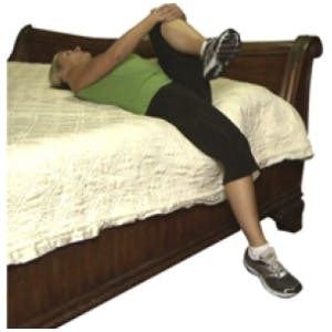
  1. Supine hip flexor stretch

  • Lie flat on your back on a bed with one leg hanging over the side
  • Pull your opposite knee tight toward your chest until a comfortable stretch is felt
  • Hold this stretch for 30 seconds
  • Complete three repetitions on each leg
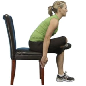
  1. Sitting piriformis stretch

  • While sitting on a chair, cross one leg over your opposite knee
  • Slowly bend your body forward until a comfortable stretch is felt
  • Hold this stretch for 30 seconds
  • Complete three repetitions on each leg
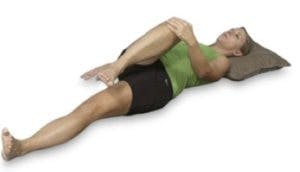
  1. Supine piriformis stretch

  • Lie on your back on the ground
  • Bend one knee up and grab it with your opposite hand
  • Pull your leg across your body, toward your shoulder, until a comfortable stretch is felt
  • Hold this stretch for 30 seconds
  • Complete three repetitions on each leg

Regularly performing these stretches will keep your hips mobile and your risk for hip pain down. But if problems do still manage to arise, it’s best to consult with a physical therapist, who will perform a comprehensive evaluation and design a personalized treatment program that’s suitable for your needs, abilities, and goals.

Full-scale Evaluation Of Various Treatments For Knee Osteoarthritis

Knee osteoarthritis: a debilitating and costly concern

Osteoarthritis (OA), a condition characterized by the breakdown of articular joint cartilage, occurs most frequently in the knee where it’s a major source of pain and disability. Patients with knee OA consequently experience an inability to perform activities of daily living and their quality of life (QoL) is also negatively affected. Today, more than 30% of adults over the age of 60 experience functional limitations due to knee OA, with health care costs related to the condition estimated at $60 billion annually. By 2020, it’s estimated that 11.6 million people will experience limitations due to the disease, making treatment of knee OA a serious priority. There are currently a number of treatments options available for knee OA (arthroscopic surgery, knee capsule injections, non-steroidal anti-inflammatory drugs [NSAIDs]) but few have proven to be as effective as physical therapy. Since the risk of knee OA is so high in aging adults, it’s imperative that physical therapists have a complete understanding of the available treatment methods and which ones are most effective. To address this concern, a systematic review was created to review current trends for treatment of knee OA and to compare the effectiveness of each intervention.

Appropriately categorizing each applicable study

A number of reputable databases were searched for studies pertinent to the focus of the review. Only studies that were peer-reviewed, published between 1996-2007 and consisting of a control group were considered for inclusion. The quality of each study was determined by the rating system of Sackett’s five levels of evidence, which allows for three grades of recommendations:

Level I: Studies consisting of randomized controlled trials (RCTs) with sample size of 100 or more subjects and low incidence of false-positive and false-negative errors

Level II: ” RCTs with sample size of less than 100 and high incidence of false-positive and false-negative errors

Level III: ” non-randomized, concurrent, cohort comparisons between subjects that did not receive treatment

Level IV: ” non-randomized, historical, cohort comparisons between current subjects who did receive treatment and former subjects who did not

Level V: ” case series without control

Recommendations were subsequently broken down using this system: grade A recommendations were supported by at least one level I study, grade B were supported by at least one level II study, and grade C were supported by any combination of levels III, IV or V studies.

Ample number of studies produces clear-cut set of recommendations

A total of 22 studies were found in the databases that were relevant to the subject matter, and after using the inclusion criteria, 15 studies made the cut and were evaluated according to Sackett’s. Of these, there were two level I trials, 12 level II trials, and two level V trials. The following recommendations were then created according to the findings of each study:

Grade A recommendations

1. Class-based exercise is more effective than home-based exercise to decrease the pain associated with knee OA.

2. True or electro acupuncture has been shown to relieve pain in patients with knee OA when compared to patients that receive sham acupuncture.

Grade B recommendations

1. Mechanical unloading using a Zuni Exercise System of patients with knee OA may not help with pain reduction.

2. The use of a neoprene brace and/or therapeutic tape on patients with knee OA may help reduce pain and regain function.

3. Patients that are treated in a clinic or classroom setting benefit more from exercise and manual therapy than those given instructions for home-based exercise.

4. Aquatic or hydrotherapy has positive effects on the strengthening of patients with knee OA.

5. Manual therapy and exercise is shown to increase quadriceps strength, while decreasing disability.

Grade C recommendations

1. An evidence-based exercise and health education program to inform patients of OA is useful in patient comprehension of the disease.

2. Stretching, strengthening exercise, and perturbations are all useful interventions for aging adults with knee OA.

In addition, there was a clear trend noticed in the studies that patients who received treatment from a physical therapist directly showed better scores on the Western Ontario and McMaster Osteoarthritis Knee Index (WOMAC) and had decreased pain when compared to those who didn’t receive treatment. This means physical therapy should be considered an effective treatment option for knee OA, particularly when these recommendations are followed. As always, future studies are needed for additional support of physical therapy’s effectiveness and to further investigate specific modalities, but for now, these recommendations should serve as a useful set of guidelines in helping physical therapists determine specific treatment programs for patients with knee OA.

-As reported in the spring ’09 edition of the Journal of Geriatric Physical Therapy

Got Back Pain? Go Conservative!

A study published in a popular orthopedic journal recommends that the simplest in most cases, to manage low back pain, is to see a physical therapist and take anti-inflammatory medications. A number of conservative research studies provide solid evidence that conservative care is the right way to go. The article stated, “Surgery should be the last option, but too often patients think of surgery as a cure all and are eager to embark on it,” Madigan says. “Also, surgeons should pay close attention to the list of contraindications, and recommend surgery only for those patients who are truly likely to benefit from it.” Read it in its entirety,  click here.


4.81

Based on 153 reviews